Top 14 Crazy Time Casinos

1Win
- Very large library of casino games and slots
- Integrated sportsbook with live betting options
- Account verification (KYC) can be slow at times

Mostbet
- Generous welcome bonus and frequent promotions
- User-friendly mobile apps for Android and iOS
- Bonus offers come with wagering requirements

1xBet
- Extremely wide range of betting markets and casino games
- Generous bonuses and multi-level VIP / loyalty program
- Complex bonus terms and high wagering requirements

Nagad88
- Combined sportsbook and casino in one platform
- Mobile-friendly site and apps for Android / iOS
- Payment delays
Dafabet
- Long-established, reputable Asian-focused betting brand
- Large portfolio of casino and live-dealer games
- Some bonuses have relatively high rollover and time limits

22Bet
- User-friendly website and mobile apps
- Supports both fiat currencies and cryptocurrencies
- Some bonuses have short validity and strict conditions


About Crazy Time
Famous Crazy Time was launched in 2020 by one of the most notable software providers so far. It features a 54-segment wheel and an opportunity to participate in lucrative bonus rounds. The maximum possible win in the game is up to 25,000x of the bet amount, which makes live Crazy Time even more exciting. With an RTP of 95.4%, the game can compete with the most popular online casino slots. It is available on both desktop and mobile devices and also supports a demo mode for practice.
| Developer | Evolution Gaming |
| Type | Live Game Show |
| Release Year | 2020 |
| RTP | 95,4% |
| Wheel | 54 Segments |
| Maximum Payout | 25,000 |
| Bonus Rounds | Cash Hunt, Coin Flip, Pachinko, Crazy Time |
| Platforms | Desktop, Mobile App |
| Demo Mode | Yes |
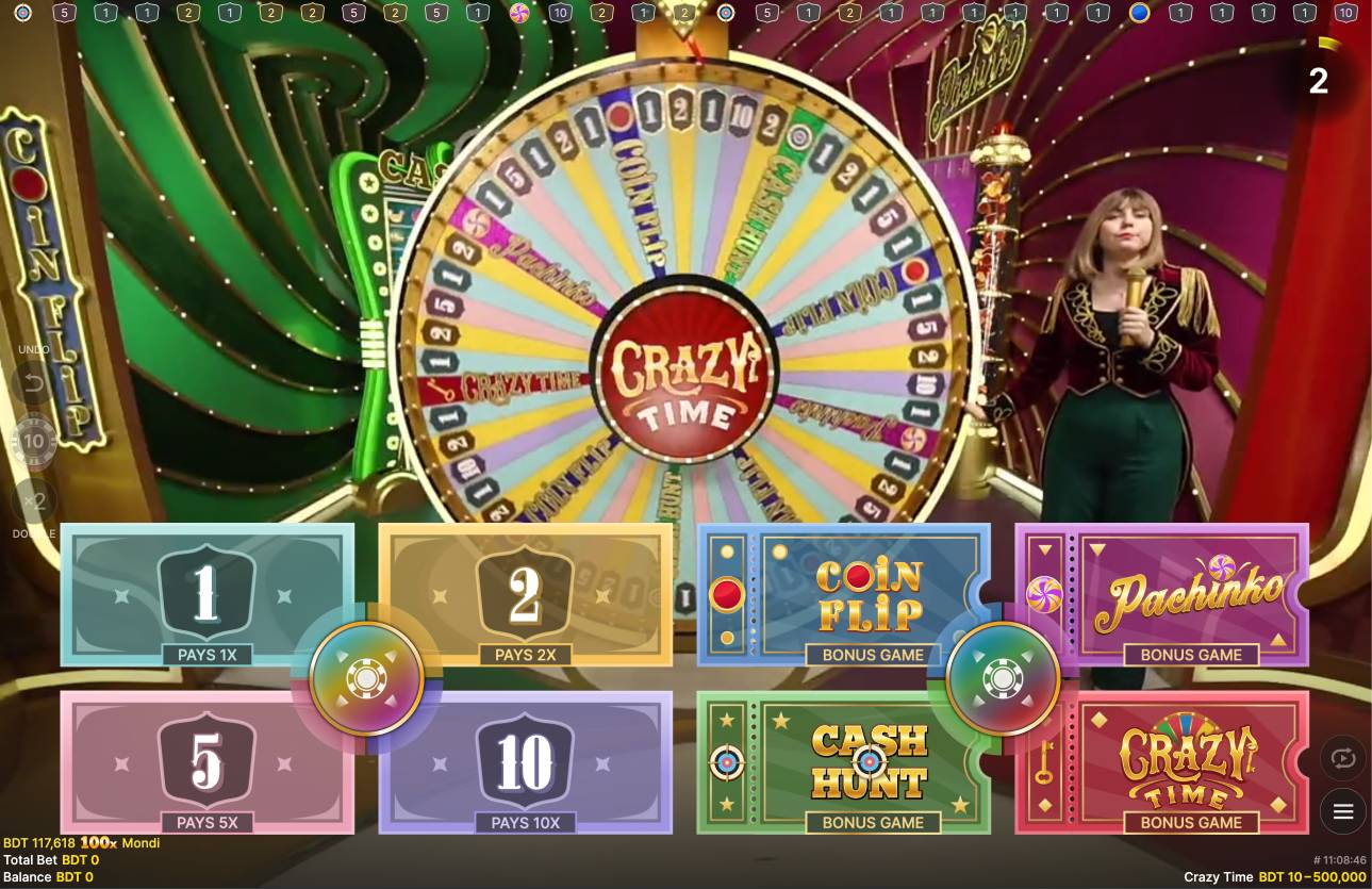
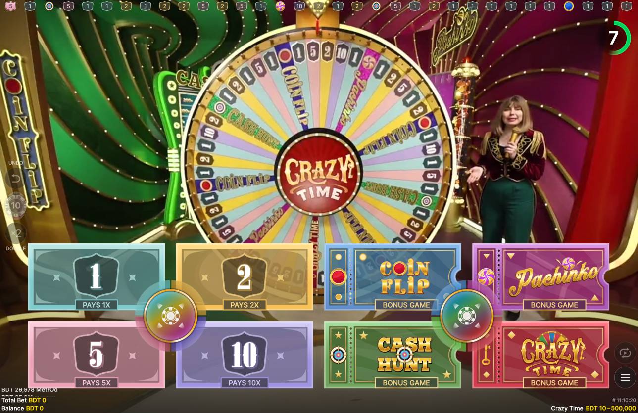
Rules of Crazy Time Live

The popular online casino game Live Crazy Time is played on the wheel with 54 sectors. When playing it, you will bet on one or several sectors, trying to predict the outcome of the round. The wheel features numerical values (1, 2, 5, and 10) as well as special bonus sections, including Coin Flip, Cash Hunt, Pachinko, and Crazy Time.
Once the bets are placed, a special dealer spins a big wheel. Meanwhile, an additional panel, called the Top Slot, rotates above the main wheel. This special mechanism defines the multiplier for each particular round. If the outcome matches the sector on which the multiplier lands and the bet is placed, the winnings are increased.
Payouts for the numbered sectors correspond to their face value. If you bet on two, then your payout will be multiplied by this value. If the wheel lands on one of the bonus sectors, an additional mini-game will begin, and you will be able to win even more money if you are lucky enough.
Bonus Games in Crazy Time

Those players who are keen on thrilling online casino games with simple rules and mechanics will definitely pay attention to Evolution Gaming Crazy Time. One of the main features of the game is that it offers four bonus rounds with lucrative rewards for those who manage to win. Each bonus game offers its own mechanics, atmosphere, and chances for serious winnings. Players can make up to 20,000x of the bet amount by playing them.
Coin Flip

In this game, you need to guess the side of the coin to win money. A random multiplier is assigned to each side before the round starts. Afterward, the coin flies upward, and the outcome becomes clear.
Most common multipliers are x5 – x20. Sometimes x100 and higher, especially with the additional bonus from the main wheel. Although this is the simplest game mechanically, it's perfect for those who value speed and minimal waiting.
Cash Hunt

One of the most popular bonuses among viewers. In this bonus game, you will have to choose your own target. The field is divided into 108 sectors with multipliers. Before choosing, all values are shuffled and hidden.
The most common multipliers range from 80 to 100x. However, players can make even bigger wins of 300-500x of the bet amount. In enhanced rounds, even higher multipliers are possible. Cash Hunt is prized for its personal touch; the outcome depends on your choices, even if everything remains random.
Pachinko

This bonus is based on the principle of Japanese slot machines, where a ball or token bounces between pins.. The host launches a disc from the top of a vertical board dotted with pins. Multipliers and "DOUBLE" fields are located below. The disc bounces chaotically and eventually lands in one of the spaces.
Base multipliers range from x5 to x100. Rare multipliers are x500 and higher. Hitting a "DOUBLE" doubles the value, and the wheel spins again. Repeated "DOUBLES" can increase the multipliers exponentially, up to x10,000.
Crazy Time

The rarest, largest, and perhaps most spectacular bonus round. It gives the entire game its name. A 64-sector wheel is offered in this special studio. Players choose colors before the round starts. The wheel begins to spin, and your chosen flapper determines the outcome. If it lands on the "DOUBLE" or "TRIPLE" sector, the values increase, and the wheel spins again (only for those who selected the corresponding color).
Players win in most cases 20-100x of the bet amount. Maximum recorded payouts are over x20,000 of the bet.
How to start playing Crazy Time?
The Crazy Time game is a very simple entertainment that requires no previous experience. Even if you are new to it, it will not take you much time to place your first bet there. Read our step-by-step guide to learn more about how to start playing the game.
Choose a reliable online casino
Select a reliable online platform from the list above.
Register on the website
After selecting a gambling site, you need to register there. Go to the website and press the sign-up button to create an account. Complete the form with all the required data.
Log in to your account
Once the signup is done, you need to log in to your account.
Add funds to your balance
Go to the Cashier or Deposit section and pick a payment method that you are going to use to transfer funds. Complete a deposit transaction to play for real money.
Find Crazy Time
Use the search bar or the Live Casino/Game Shows section to find game and launch it.
Place a bet
You can choose to bet on one of the number sectors (1, 2, 5, 10) or on the bonus rounds: Coin Flip, Cash Hunt, Pachinko, and Crazy Time.
Enjoy the game
The dealer spins the wheel live, and everything depends on luck. Just watch and enjoy the process.
RTP and multipliers in Crazy Time

40.17%
646 Lands

23.2%
373 Lands

12.87%
207 Lands

6.9%
111 Lands

3.48%
56 Lands

3.23%
52 Lands

8.08%
130 Lands

2.05%
33 Lands
The RTP (Return to Player) varies depending on what you bet on. The highest RTP is for bets on numbers, especially 1 and 2, as they appear more often and bring consistent, but modest, winnings. The RTP here is around 96%. At the same time, bonus round bets (such as Cash Hunt or Crazy Time) have a slightly lower RTP, from 94% to 95%.
This is because they occur less frequently, but offer the chance for big wins thanks to powerful multipliers. Coin Flip and Cash Hunt can multiply your bet by 500x, while Pachinko can multiply it up to 10,000x. The highest potential payout in Crazy Time is a 20,000x multiplier, available in the bonus round of the same name if you successfully match the Top Slot. Understanding the RTP structure and probabilities helps you develop a conscious strategy: either play carefully with frequent but small payouts, or take risks for big wins in bonus games.
| Wheel segment | Number of segments | Chance of getting | RTP | Average WIN |
|---|---|---|---|---|
| Number 1 | 21 | 38.89% | 97.68% | 1:1 |
| Number 2 | 13 | 24.07% | 96.55% | 2:1 |
| Number 5 | 7 | 12.96% | 94.58% | 5:1 |
| Number 10 | 4 | 7.41% | 93.83% | 10:1 |
| Coin Flip | 4 | 7.41% | 95.70% | 13x, up to 5,000x |
| Cash Hunt | 2 | 3.70% | 94.33% | 25x, up to 12,500x |
| Pachinko | 2 | 3.70% | 94.33% | 25x, up to 5,600x |
| Crazy Time | 1 | 1.85% | 94.41% | 50x, up to 8,000x |
Crazy Time App
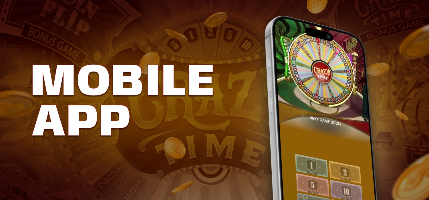
Almost every platform provides its own gaming app that allows you to play via a mobile device. Below you will find a guide on how to download the Crazy Time app:
Installation on Android: Quick and Easy
To start playing Crazy Time on Android, follow these steps:
- Open the website of your chosen casino on your smartphone's browser.
- Go to the mobile app section.
- Download the APK file by following the instructions on the website.
- In your phone's settings, allow installation from unknown sources.
After installation, open the app, log in to your account, and you're ready to play.
How to Install the App on iOS
If you use an iPhone or iPad, installing Crazy Time is even easier, especially if the casino has the app available in the App Store:
- Go to the casino's website from your device.
- Find the download link in the App Store.
- Confirm the installation as usual, using Face ID or your password.
- After installation, open the app, log in to your account, and start playing.
If the app is unavailable, you can always use the mobile version of the website; it runs smoothly, loads quickly, and is just as good as the app.
Crazy Time Demo

If you're just starting out with Crazy Time or want to better understand how the game works, the demo mode is a great way to try everything out risk-free. In some online casinos, you can find a so-called preview mode, where you can watch the game flow, but without participating, the rounds unfold, and the wheel spins without having to wager real money.
It's important to understand that there's no full-fledged demo in the traditional sense, as Crazy Time is a live game with a real dealer. However, you can try the game as a guest or without a deposit, just to see how it works.
Screenshots of Crazy Time


Legality of Crazy Time
The legality of playing Crazy Time Bangladesh directly depends on the platform you use to play. Crazy Time was developed by Evolution Gaming, a regulated and known company in the world of gambling. On the other hand, if you are playing the game via the online casino, you can do it without any limits, as you will play on the offshore platform in this case, which is not prohibited by local legislation.
Reviews from Players about Crazy Time
Abdur Maroof
"Crazy Time didn't hook me right away, but it's now my favorite live casino game. You really get the TV show vibe. The most memorable moments were when the bonuses landed, especially Pachinko. I once hit a 200x multiplier that was unforgettable."

Ayesha Siddiqua
"I'd never played in a casino before, but the design and hosts at Crazy Time made an impression. It's more of a fun experience than a typical gambling game. Sometimes I win small amounts, but I play for the thrill, not the money."

Shamima Sultana
"Sometimes the game is enjoyable, and other times it's addictive waiting for bonuses. There was a period when the bonus didn't hit for almost 30 spins; it was tiring. But when it does, it feels like you've hit the jackpot. The main thing is to know how to stop."

Fatema Khatun
"Pleasant visuals, good hosts, and fairly clear rules. Crazy Time is suitable even for those new to casinos. I bet cautiously, but I've already had some nice payouts. Coin Flip gave me 80x, and I was thrilled."

Mohammad Rahman
"I've tried many games in my life, but I've never seen such a combination of showmanship and randomness. Sometimes it seems like winnings are too rare, but the thrill compensates for that. The main thing is to know when to quit and not chase every bet."

Arif Ahmed
"The game is suitable for those who want to relax without getting bogged down in complex strategies. I play a couple of times a week, bet small amounts, and overall I'm happy. My last Cash Hunt win was 75x, not a million, of course, but still nice."
Pros and Cons of Crazy Time
The game has both positives and negatives, with advantages outweighing some minor negatives. This game is popular with many, and even beginners can make some winnings. Below is a balanced look at its strengths and weaknesses.
- Well-designed format. The game of Crazy Time is not another live casino software, as it combines the thrill of a famous TV Show. Thanks to live hosts and a well-thought-out presentation, sessions are quick and don't become boring even after extended play
- Availability of bonus rounds. Apart from standard gameplay, Crazy Time offers extended mini-games where you can win big prizes if you are lucky enough
- Potentially High Wins. Players can win hundreds of times their bet amount. The luckiest gamblers participate in bonus rounds with an opportunity to catch up to 20,000 of the bet amount
- Low entry barrier. Playing is possible even with a small budget. This is especially important for beginners or those who simply want to test the game without serious risk
- Presence and live interaction. The live stream format and the ability to watch the game in real time increase engagement. Moreover
- Randomness. The player has virtually no influence on the outcome. Unlike poker or roulette with a betting system, here you can only watch and hope for luck
- Infrequent bonus games. Some sessions go without bonuses for dozens of rounds in a row. This reduces the momentum, especially if the player relies specifically on bonus potential
- Volatility. While large wins are possible, it's just as easy to quickly go into the red. Comfortable play requires discipline and budget control
- Technical nuances. Due to the live game format, the quality of the broadcast directly depends on the stability of the internet. Delays, interruptions, and freezes are common, especially when playing on mobile devices
Frequently Asked Questions about Crazy Time
What is Crazy Time?
This is an interactive game with a real host, developed by Evolution. Players place bets on sectors of a large wheel, which features both numbers and bonus rounds. The mechanics themselves are reminiscent of a television game show with audience participation.
Can I play on a phone or tablet?
Yes, the game is fully compatible with mobile devices. It runs in a browser and is also available through mobile apps for Android and iOS, provided the casino offers this feature.
Where can I see game results?
The latest results are displayed directly in the game interface. Additionally, some platforms publish advanced statistics, such as bonus frequency, odds, and round history, which are useful for analysis and betting planning.
Is there a free mode?
Some online casinos offer a demo version of Crazy Time, but a full demo mode is rare due to the live streaming format. However, you can learn the mechanics through recorded rounds or tutorials.
How do I start playing?
First, choose a reputable, licensed casino with the Evolution provider. Then, register, verify your account (if necessary), top up your balance, and find the game in the live dealer section. After this, you can start placing bets.